Copyright © 2007-2018 Russ Dewey
Subcortical Structures and Functions
So far we have examined mostly the large, visible areas on the surface of the brain: the cerebral cortex. While this is an important part of the brain, it is only the surface. Below the cerebral cortex are other structures, called subcortical (literally "below the cortex") structures.
You need not memorize most of these structures for an introductory psychology class. The main idea is that there are many specialized areas below the cortex. A few of them, such as the limbic system and amygdala, are frequently referenced in psychology articles.
The following table lists the most commonly accepted, classic functions attributed to brain areas below the cerebral cortex. This list is greatly oversimplified. Exceptions, controversies, and complications are left out.
FOREBRAIN
(below cerebrum)
Basal ganglia
–involved in motor control
Limbic system
–the limbic system is not one structure but several: hippocampus, amygdala, septum, cingulate gyrus, and others
–the middle of the septum is one location of "pleasure centers"
–the amygdala involved in emotions, fear, and defensive reactions to external threats
–the hippocampus interacts with temporal lobe to help establish event memory
Thalamus
–looks like two eggs or small, joined footballs
–implicated in control of sleep and attention
–a relay station; receives input from eyes, ear, spinal cord, relays information to cerebral cortex
Hypothalamus
–involved with basic functions like eating, sex, temperature control, sleep, aggression
–produces sex, growth and stress–related hormones carried down axons to pituitary gland, released from there into bloodstream to activate and organize distant body systems
MIDBRAIN
Tectum
–consists of four bumps, the colliculi
–superior (higher up) colliculi related to eye movement and localization of objects
–inferior (lower down) colliculi related to sense of hearing
Tegmentum
–includes red nucleus and substantia nigra, involved in control of movement
–contains part of reticular formation (see below)
HINDBRAIN
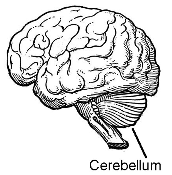
Cerebellum
–appears as a separate structure with a cauliflower–like appearance behind the brain
–controls fine motor movement, timing; motor memory, planning of movements, plus practice–related memory and detection of errors in non–motor tasks
Reticular formation
–consists of densely packed, reticulated (netlike) cells located in central core of hindbrain
–thought to activate thalamus and cortex, therefore often called the "reticular activating system"
Pons
–connects cerebrum with cerebellum
–contains centers regulating sleep, feeding and facial expression
Medulla
–first part of the brain above the spinal cord, essentially a continuation of the cord
–controls heart and respiration rate, digestive functions, blood pressure
The brain stem is the part of the nervous system starting where the spinal cord enters the brain, at the base of the skull. The brain stem (not the cortex!) is the "core" of the brain.
The brain stem extends upward to the hypothalamus. Strictly speaking, every structure on the above list except the cerebellum is part of the brain stem.
However, the term brain stem is used most commonly to refer to part of the brain that appears as a continuation of the spinal cord into the brain: the hindbrain and lower portion of the midbrain.
What is the brain stem? What effect can be produced by small injuries there?
The brain stem controls vital functions such as breathing and temperature regulation. Even tiny injuries to the brain stem can produce coma (long-lasting unconsciousness) or death.
My wife worked in an Emergency Room, and she remembers a woman being brought by ambulance after an automobile accident, unconscious, with pupils fixed and dilated and unresponsive to light, indicating brain death. The only injury doctors could find was a small puncture wound to the brain stem.
The Cerebellum
The cerebellum is part of the hindbrain, but it is not part of the brain stem. The word cerebellum means little brain. The cerebellum appears as a lobe at the back of the brain with convolutions smaller and denser than the cerebrum.

Among its other duties, the cerebellum serves as a storehouse for motor memories: memories of activity sequences. When the cerebellum is damaged from a birth defect or injury, the common result is loss of fine motor coordination called spasticity. The spastic gait is a distinctive way of walking in which first one foot then the other is laboriously set forward. It is a sign of cerebellar damage.
Where is the cerebellum, and what are its functions?
What happens when the cerebellum is damaged?
The cerebellum also participates in error-correction and screening out incorrect responses by other brain systems. This was a surprise to neuroscientists who initially thought the cerebellum was only involved in motor control.
The cerebellum contains spectacular cells called Purkinje cells, named after a Czech physiologist. His name (POOR-kin-yay) is usually mispronounced "per-KIN-gee" in the United States.
Packed around the Purkinje cells in the cerebellum are smaller neurons called granule cells. They are called "granule" cells because they are so tiny they appeared as little particles (granules) when first discovered.
Granule cells turned out to be complete nerve cells. There are as many as 40 billion granule cells in the cerebellum.
How many neurons are in the brain, according to modern estimates?
Shepherd (1974) wrote, "It is commonly stated that the human brain contains 10 billion nerve cells, as evidence of its fantastic complexity; clearly those statements do not take into account the granule cells of the cerebellum!" Humans are, as Shepherd put it, "very cerebellar animals."
The Peripheral Nervous System
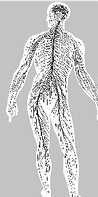
The peripheral nervous system (PNS) consists of nerves outside the brain and spinal cord: those which innervate (supply with nerves) the muscles and glands.
There are two major subdivisions of the PNS: the somatic nervous system and the autonomic nervous system. Nerves controlling the somatic and autonomic systems often begin within the central nervous system but have most of their effect outside it.
What are two major subdivisions of the peripheral nervous system?

The somatic nervous system controls skeletal muscles. These are the muscles we use for moving our limbs. Skeletal muscles are attached to the bones of the body, which is how they got their name. Soma (the root of the word "somatic") means body in Latin. So the somatic (body) nervous system controls skeletal muscles (attached to bones of the body).
What is controlled by the somatic nervous system?
The autonomic nervous system controls smooth muscles. Smooth muscles make up the walls of the intestine, glands, and blood vessels. Autonomic means automatic or capable of acting on its own. The glands and intestines seem to act on their own, although there are some exceptions.
For example, some actors can make tears flow voluntarily. Tears come from tear ducts, which are exocrine glands. Like all glands, they are innervated by the autonomic nervous system. This is the exception that proves (tests) the rule, because actors typically accomplish this feat by vivid imagination. They fool the autonomic system into reacting as if a situation is real.
What does "autonomic" mean? How are voluntary tears an exception to the general rule?
The autonomic nervous system itself has two subdivisions with opposite effects. The sympathetic part of the autonomic system swings into action during fight or flight.. Your adrenaline flows, your heart rate and breathing rate go up, your palms sweat, and your skin may grow clammy as blood flows away from skin to skeletal muscles.
Sympathetic nervous system activation also leads to an excited mental state in which subtle, complex memories may become unavailable. An example is "blanking" on a test. Our sympathetic nervous systems were built to take action, not tests.
Which part of the autonomic nervous system produces the "fight or flight" reaction?
The parasympathetic system takes over as your body returns to a period of rest and relaxation. It diverts your blood supply from skeletal muscles back to your intestine so you can resume digestion of food.
The parasympathetic system also slows your heart rate and respiration rate, and it allows you to replenish your system with energy. Your skin warms and a rosy glow returns to your cheeks.
What are effects of parasympathetic activation?
Many people find that digestive functions are inhibited while they are traveling or at school. Then, when they get home, they head for the bathroom. The nervous system has switched from sympathetic nervous system arousal, keeping the person alert and ready for stress, to parasympathetic arousal, allowing rest and recovery.
---------------------
Reference:
Shepherd G. M. (1974). The Synaptic Organization of the Brain. New York: Oxford University Press.
Write to Dr. Dewey at psywww@gmail.com.
Don't see what you need? Psych Web has over 1,000 pages, so it may be elsewhere on the site. Do a site-specific Google search using the box below.
