Copyright © 2007-2018 Russ Dewey
The Visual System
The visual system is studied more than the other perceptual systems in humans. Humans (like other primates) are dependent upon vision and have a large portion of the brains devoted to sight.
The sense organ for sight is the eyeball. It gathers light into a focused image on the back of the eye. Focusing is done by refracting (bending) light as it passes through the transparent tissues and fluids of the eyeball.
Refraction is familiar to anyone who has held a magnifying glass under the sun. Light from the sun is bent by the lens, concentrating light on a small spot where it becomes hot enough to start a fire.
The curvature of the eye performs a similar function. That is why it is dangerous to look directly at the sun: it can burn the back of your eye.
Why is it dangerous to look directly at the sun?
A magnifying glass creates a point of focus because its image is dominated by a single source of light, the sun. Light from the environment creates a focused image in our eyes because light comes in from every direction. Each point in the environment is focused on a different point on the back of the eye.
The back of the eye receives an inverted (upside-down) image of the world. Descartes discovered the inverted image on the back of the eyeball in 1637.
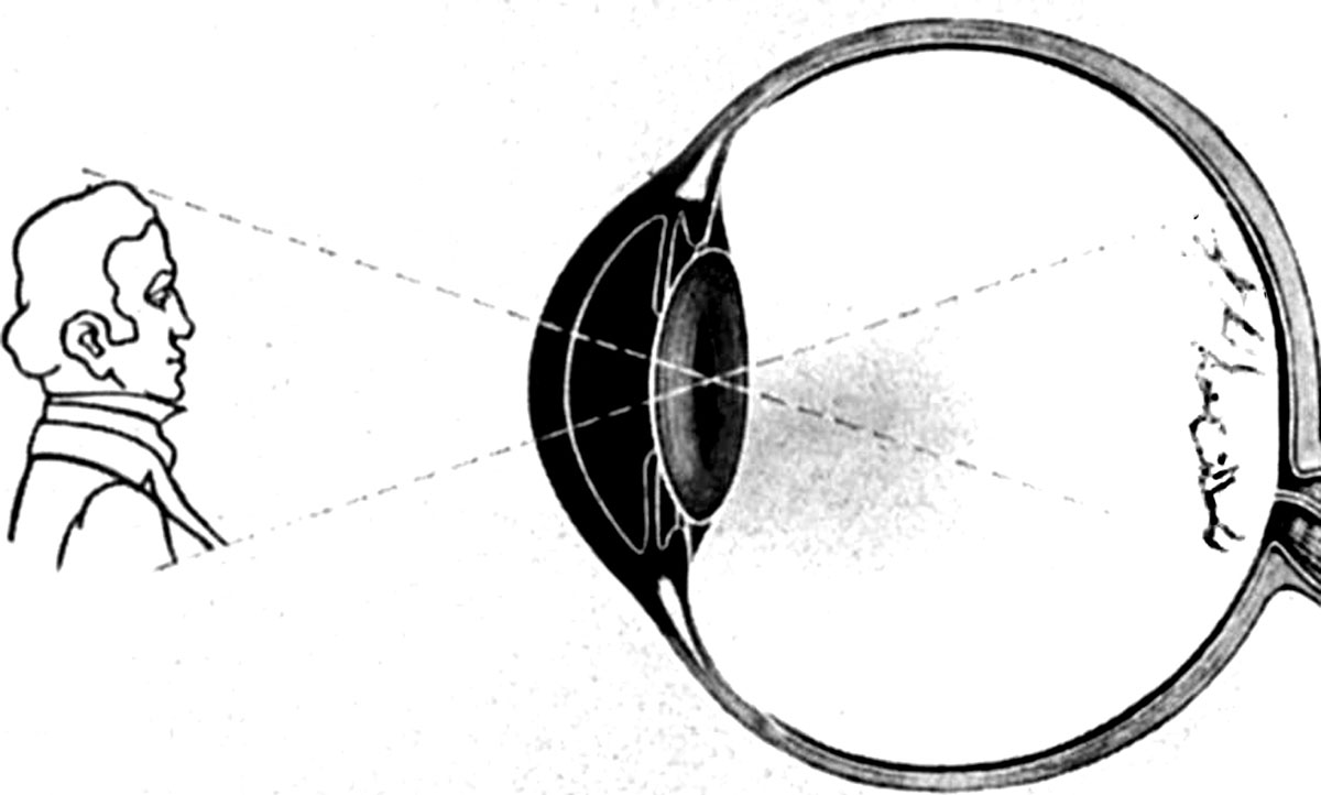
The back of the eye receives an inverted image
Descartes scraped tissue off the back of an eye and inserted a small thin piece of paper in its place. Holding up the eye, he observed a small, sharp, upside-down image of the environment on the paper.
How did Descartes observe the inverted image on the back of the eye?
Structures of the Eye
The frontmost layer of the eye is the cornea. It is a tough, clear covering, bathed in tears. It is very painful when scratched.
We blink in order to keep the cornea moist. The cornea refracts light, so it is the first stage in the process of focusing the visual image.
The transparent liquid behind the cornea, called the aqueous humor, also refracts light and helps bring the image of the outside world closer to a focus. Behind the chamber containing the aqueous humor is the iris, the colored part of the eye.
The color of the iris (and thus the eyes) is determined by the amount of pigment contained in them. Albinos, who lack pigment, have pink irises.
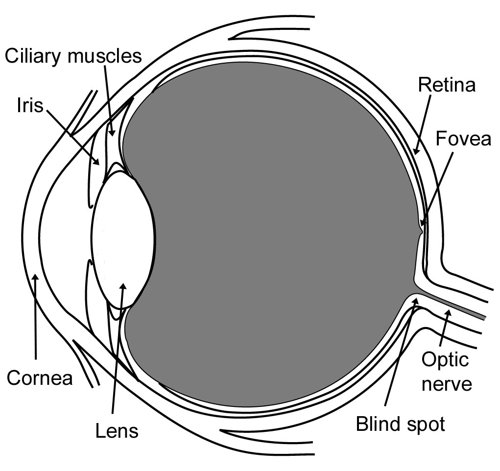
Structures of the eye
The iris has a complex internal structure, including a set of muscles all its own. The muscles allow the iris to change the size of the hole in the middle of the iris, called the pupil.
Pupil size changes to compensate for changing levels of light: this is the pupillary reflex. It regulates the amount of light entering the eye. If you are in a brightly lit room, your pupil will shrink or constrict.
If you are in a dimly lit room, your pupils will enlarge or dilate. The fully dilated pupil has 17 times greater area than the fully constricted pupil.
What is the cornea? The iris? What is the pupillary reflex, and what is its function?
Muscles in the iris are controlled by the brain stem. The pupillary reflex works only as long as the brain stem is alive.
Doctors check the pupillary response of unconscious people brought into an Emergency Room. If the pupil is fixed and dilated, wide open and unresponsive to light, the patient is dead.
Why are fixed and dilated pupils a bad sign, in the Emergency Room?
The Lens and Visual Accommodation
Immediately behind the iris is the lens. Shaped like a magnifying glass, it refracts light even more than the cornea and aqueous humor.
The lens plays a primary role in focusing the visual image on the back of the eye. The lens is not rigid; it can be bulged and flattened somewhat. The lens changes its shape when pulled by fibers called the suspensory ligaments attached to the ciliary muscles.
When you focus on near objects, you strain the ciliary muscles. They cause the lens to bulge, which focuses the eye on nearby things. The ciliary muscles relax when you look at something far away.
What is the primary role of the lens? Why is it a strain to focus on close objects?
This adjustment of lens shape is called visual accommodation (a-com-o-DAY-tion). Researchers have found that even when people imagine looking at faraway objects, their ciliary muscles relax, showing visual accommodation.
What is visual accommodation?
Near-sightedness (myopia ) and far-sightedness (hyperopia or hypermetropia) are common problems of vision occuring when the image from the environment is not focused correctly on the back of the eye.
The line labeled focal plane in the following figure is the place where the image is clear, not blurry. Glasses and contact lenses change refraction slightly to align the focal plane with the back of the eye. That is why they are called corrective lenses.
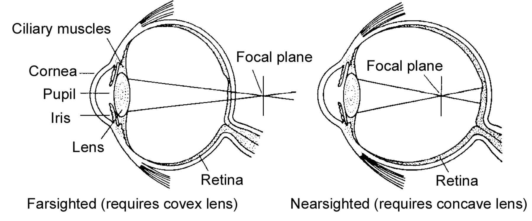
Corrective lenses are necessary if the eye is too long or too short so that the focal plane misses the retina.
After age 40 the lens of the eye becomes less flexible. Bi-focals (lenses with different focal length on the top and bottom) may become necessary, to change focus between far and near objects. This is called presbyopia (old eyes).
How do glasses and contact lenses and bifocals work? What is presbyopia?
After light passes through the lens, it goes through the chamber in the middle of the eye. It contains a gelatin-like substance called vitreous humor. Once light passes through the vitreous humor, it lands on the back of the eye, the retina (Latin for net).
The Retina
The retina consists of many layers of nerve cells. It is sometimes called a peripheral brain because it contains millions of neurons, with over 50 cell types and hundreds of millions of individual cells. They do sophisticated processing on visual information before nerve impulses are sent to the brain.
Light passes through the blood vessels and several layers of nerve cells before arriving at the photoreceptors, the rods and cones. The rods and cones are receptor cells of the eye.
As you can see from the diagram, the names are appropriate. Cones are the cone-shaped cells at the bottom; rods are the rod-shaped cells next to them. The diagram is highly simplified, showing only a few cell types.
Of what does the retina consist?
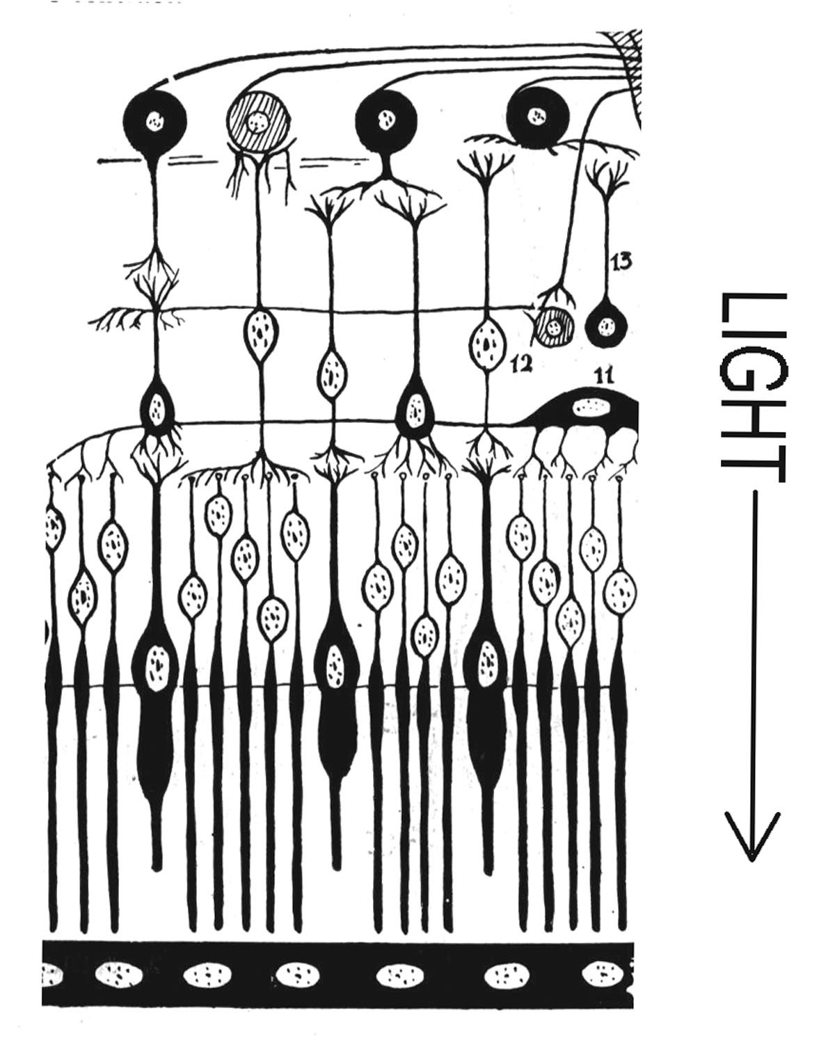
Simplified cross-section of the retina. Light enters from above.
Rods and cones consist of layer upon layer of folded cell membranes. In the following diagram, a tiny segment of a rod receptor is magnified to show the layers.
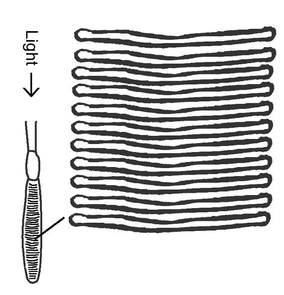
Inside rods and cones are layers of folded membrane.
Each layer contains light-sensitive chemicals. When light strikes the chemicals, it provokes a chemical reaction, generating a tiny electrochemical current that influences nerve cells near the receptors.
How do the rods and cones work?
Although rods and cones both use layers of chemicals that react to light, they use different chemicals, so they respond differently to light. Cones are specialized for color vision. They are called chromatic receptors, the root chroma- meaning color.
Rods, by contrast, are specialized for black-and-white vision. They are called achromatic (no color) receptors.
What are the eye's chromatic and achromatic receptors?
There are two other important differences between the rods and cones: (1) rods have greater sensitivity to light and (2) cones have greater acuity (ability to pick out details).
Because rods are more sensitive to light, they are the receptors we use at night. When you see objects outdoors by the light of the moon, there is not enough light to activate the cones, so you use primarily rod vision. This is what gives the nighttime world a black-and-white (achromatic) appearance.
However, our acuity is not as good at night. We cannot see fine details as easily at night, because the cone receptors are not as active in the darkness.
What are two other differences between rod and cone vision?
When light is not present, the rods and cones build up their supplies of photosensitive chemicals. This process is called dark adaptation.
Dark adaptation makes our eyes much more sensitive to light. The rod receptors become increasingly light sensitive for about 30 minutes after being deprived of high light levels
When rods are fully dark adapted, they are 10,000 times more sensitive than they are when they are bleached by strong light. This is why dark adaptation has such a noticeable effect in places like movie theaters.
How long does it take to become fully dark adapted?
People coming into a movie theater from bright outside daylight stumble around in the semi-darkness. People who have been sitting in the theater for a long time can see clearly, because their eyes are adjusted to the darkness and much more sensitive to light.
The Fovea
Rods and cones differ in their placement within the eye. Cones are concentrated in the center of the retina in an area called the fovea (Latin for fireplace).
In the fovea centralis (center of the fovea) there are no rod receptors. In the fovea, 50,000 cones fit in an area the size of a dot on the letter i.
When you look directly at something, the center part of image falls on the fovea. Only a small part falls on the fovea centralis. A thumbnail held at arm's length is sufficient to cover the fovea centralis.
What is the fovea? What is the fovea centralis? How can you create a visual stimulus that just barely covers the fovea centralis?
Near the fovea, blood vessels and other structures are less numerous, so it is easier for the light to get through. This is shown in the figure, which shows a cross-section through the fovea.
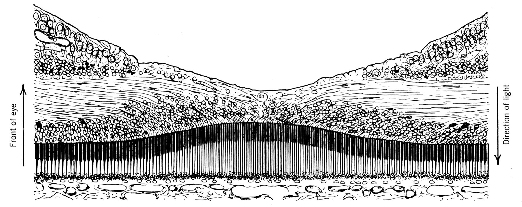
Cross-section of the fovea
The area outside the fovea is called the periphery of the retina. A thing seen "out of the corner of the eye" is casting its image on the periphery.
There are more rods in the periphery. Also, rods are more sensitive to light than cones. Therefore, one can see a faint star in the night sky more clearly if one does not look directly at it.
Amateur astronomers using light telescopes become skilled with peripheral vision. They can look sideways into a telescope, using rod vision, to detect more stars.
What is the periphery? Why can you see a dim star better in your periphery?
As a rule, rod-based vision is not very good at picking out details. That is why night vision lacks detail. It is predominantly rod vision.
Because cones are far more numerous in the fovea, acuity (the ability to pick out detail) is much greater in the fovea, compared to the periphery. However, cells in the periphery of the retina are very sensitive to movement.
Which receptors are more associated with high acuity? With movement detection?
Movements in peripheral vision trigger an eye movement automatically, using a visual circuit that runs through the superior colliculi of the brain. The eye movement centers a possible target (the source of movement) on the fovea, so it can be analyzed further.
Write to Dr. Dewey at psywww@gmail.com.
Don't see what you need? Psych Web has over 1,000 pages, so it may be elsewhere on the site. Do a site-specific Google search using the box below.
